Assessment of Myocardial Viability Using Nuclear Medicine Imaging in Dextrocardia
Por um escritor misterioso
Last updated 20 setembro 2024
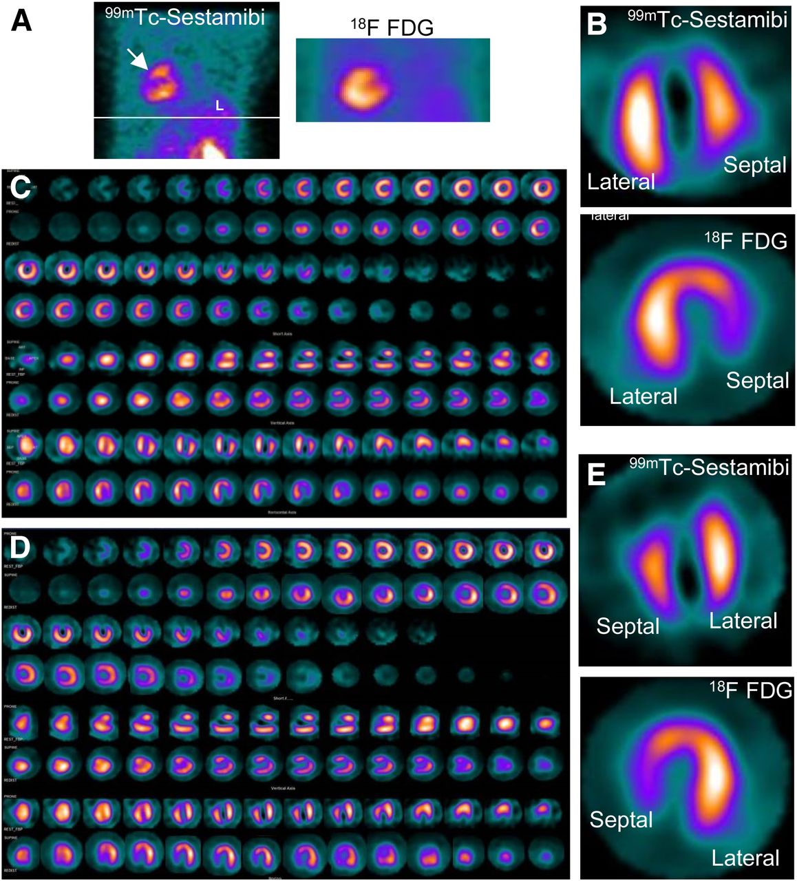
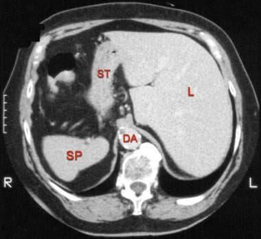
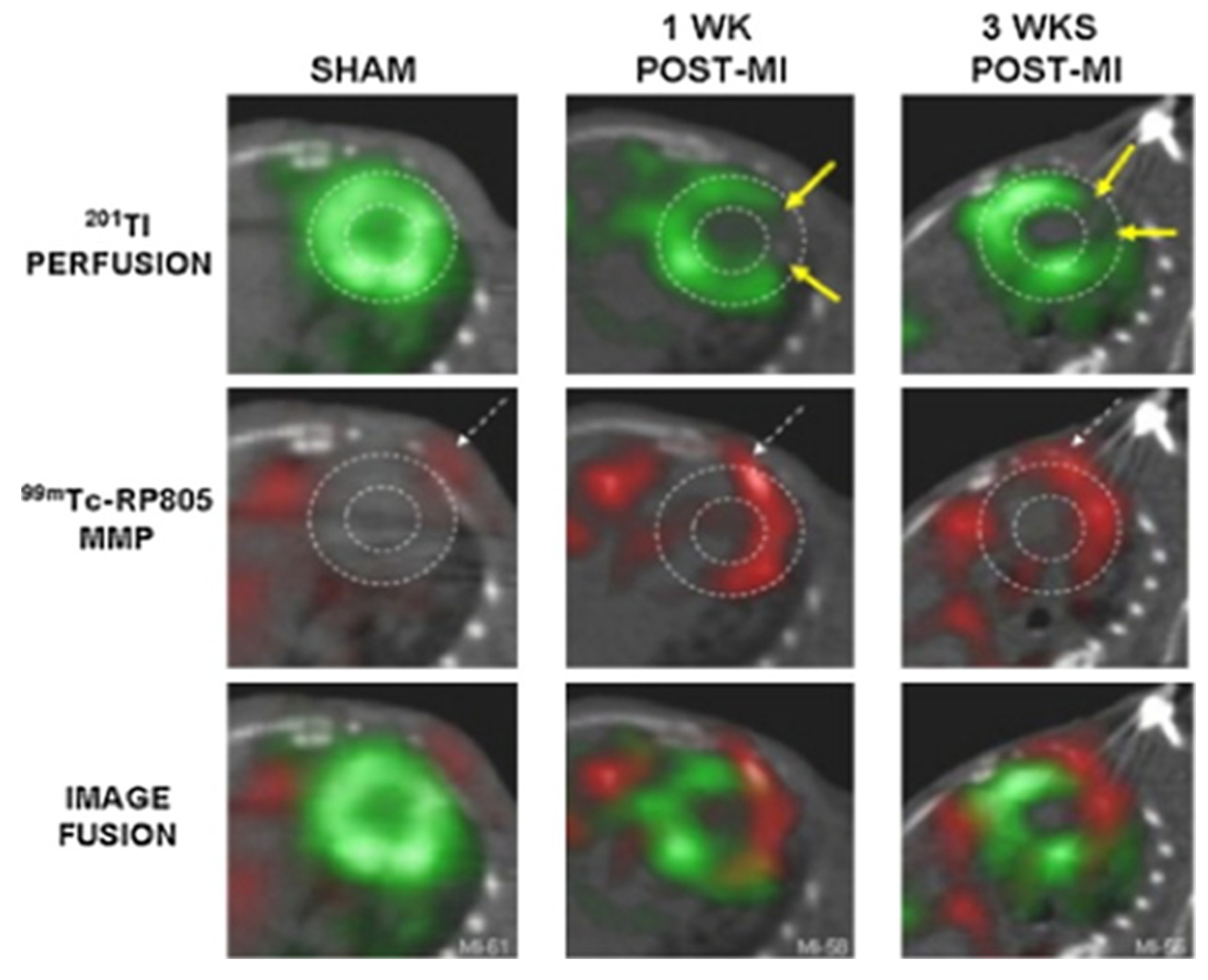
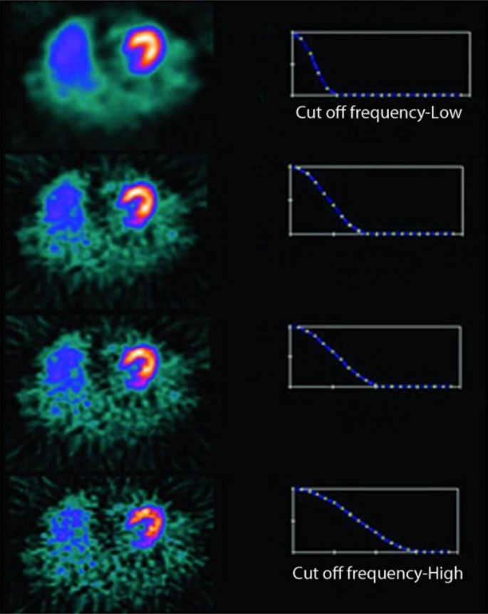
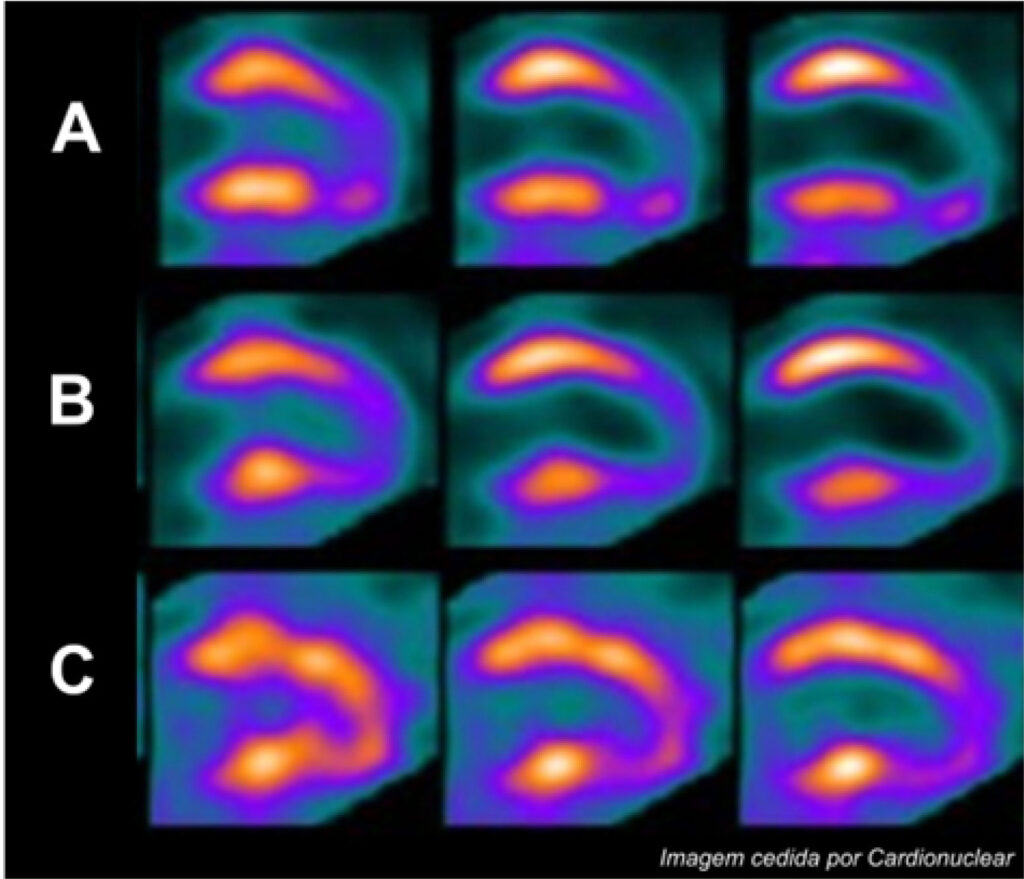
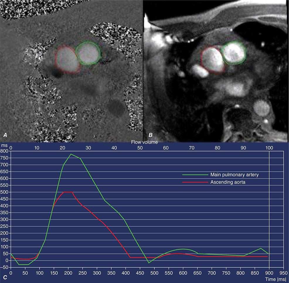
Imaging of dextrocardia in humans requires an understanding of the orientation of the heart chambers and walls. There are many types of cardiac malpositioning, such as dextrocardia (with or without situs inversus), mesocardia, and levocardia. Myocardial perfusion scintigraphy of dextrocardia has been explained in case reports and imaging atlases; however, myocardial viability assessment using nuclear medicine imaging techniques is less documented in the literature. Methods: In 2 cases of dextrocardia with situs inversus and 1 case of mesocardia, myocardial viability was assessed using 99mTc-sestamibi rest perfusion scintigraphy and 18F-FDG PET. Cardiac SPECT images of dextrocardia with situs inversus were acquired using the feet-first supine position with a 180° arc from left anterior oblique to right posterior oblique, whereas a right-lateral–to–left-lateral arc was used for mesocardia. The processing and reconstruction were done by entering the dataset for the feet-first supine position and repeating after entering the dataset for the feet-first prone position. The 2 sets of reconstructed images were compared for orientation of walls and cardiac chambers. Results: The first processing, using the feet-first supine position, revealed an interchanged septum and lateral wall in reconstructed images of dextrocardia with situs inversus. This interchange was corrected by changing the position to prone during processing of the rest perfusion and PET raw data. The display of cardiac slices in various axes matched the conventional nomenclature for the septum and lateral wall, leading to easy interpretation. However, this change was not required in the mesocardia, for which the location of the heart chambers was not interchanged. Conclusion: Because the acquisition protocol for SPECT is a semicircular orbit, the various types of dextrocardia require careful selection of the arc, with the patient positioning kept feet-first supine. Processing and reconstruction of data by changing the patient position to prone was found to be most useful method of matching the septum and lateral wall orientation for interpretation of images.
Situs inversus totalis

Tl-201 treadmill stress/rest myocardial perfusion SPECT short axis
Nuclear Medicine Imaging of Myocardial Viability

Approach to Dextrocardia in Adults: Review
SPECT myocardial perfusion imaging in patients with Dextrocardia

PDF] Myocardial Perfusion SPECT Imaging in Dextrocardia with Situs
Situs Inversus Imaging: Practice Essentials, Radiography, Computed
Pharmaceuticals, Free Full-Text

The Impact of the Coronavirus Disease 2019 Pandemic on the
Single Photon Emission Computed Tomography (SPECT) Myocardial
Assessing Myocardial Viability in Clinical Practice - ABC Imaging
Noninvasive Cardiac Imaging: Echocardiography, Nuclear Cardiology
Myocardial perfusion single photon computed tomography: An Atlas

PDF] Artifacts and pitfalls in myocardial perfusion imaging
Table of Contents — December 01, 2020, 48 (4)
Recomendado para você
-
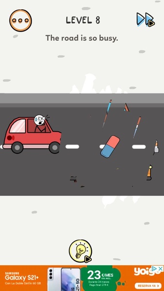
brain test level 372|TikTok Search20 setembro 2024
-
 Easy Game Brain Test Level 372 Finish shopping.20 setembro 2024
 -
Erase Puzzle for Android - Download the APK from Uptodown20 setembro 2024
-
 A Genetic Screen Implicates miRNA-372 and miRNA-373 As Oncogenes in Testicular Germ Cell Tumors: Cell20 setembro 2024
 -
 Draw Bridge Stickman Car Game on the App Store20 setembro 2024
 -
 A sense of self20 setembro 2024
 -
 Ischemic Optic Neuropathies20 setembro 2024
 -
 The answer to level 371, 372, 373, 374, 375, 376, 377, 378, 379 and 380 is DOP 2: Delete One Part - Brain Game Master20 setembro 2024
 -
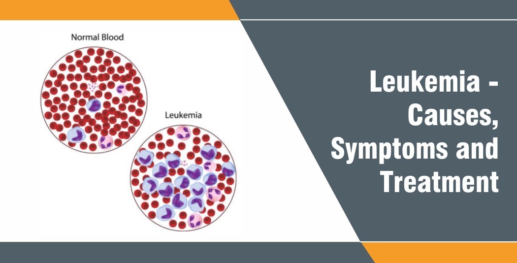 Blood Cancer - Causes, Symptoms and Treatment - Rela Hospital20 setembro 2024
 -
 Lengkap Ada Video, Brain Test Level 372 Selamatkan Kapal Titanic! ✓20 setembro 2024

você pode gostar
-
 When the story ends (shallot's story not Dragonball) how/what do you want the devs to do next? : r/DragonballLegends20 setembro 2024
 -
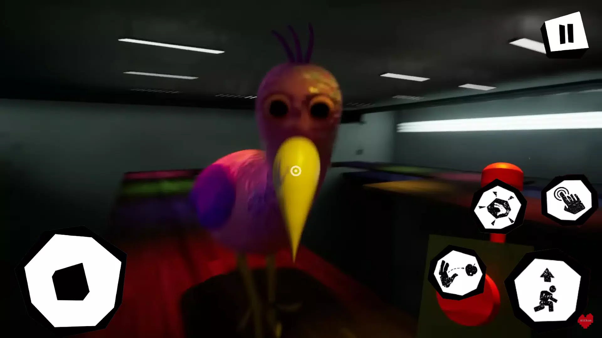 Download do APK de Garten of Banban chapter 2 para Android20 setembro 2024
 -
 Create interactive charts to tell your story more effectively20 setembro 2024
 -
 Samsung Brings Galaxy to More People: Introducing Galaxy S10 Lite20 setembro 2024
 -
 Vestido Alemã Xadrez preto e branco e veludo preto luxuoso - Princesa Urbana - Viva o Encanto20 setembro 2024
 -
 The Best Nintendo Switch Lite Games for 202020 setembro 2024
 -
 Silent Hill 2: Restless Dreams Remake (Concept Art) : r/silenthill20 setembro 2024
 -
 Read One Piece - Digital Colored Comics Vol.12 Chapter 103: Whale20 setembro 2024
 -
 Rap do Kakashi, Naruto, Sasuke e Sakura - TIME 720 setembro 2024
 -
 Is there any way to get a picture of a shirt as shown in the catalog from the website? - Scripting Support - Developer Forum20 setembro 2024