PDF] Brain Tumor Segmentation of MRI Images Using Processed Image Driven U-Net Architecture
Por um escritor misterioso
Last updated 22 setembro 2024
![PDF] Brain Tumor Segmentation of MRI Images Using Processed Image Driven U-Net Architecture](https://d3i71xaburhd42.cloudfront.net/c750894747d2b3f841de55922b2b68794295de27/7-Table3-1.png)
A fully automatic methodology to handle the task of segmentation of gliomas in pre-operative MRI scans is developed using a U-Net-based deep learning model that reached high-performance accuracy on the BraTS 2018 training, validation, as well as testing dataset. Brain tumor segmentation seeks to separate healthy tissue from tumorous regions. This is an essential step in diagnosis and treatment planning to maximize the likelihood of successful treatment. Magnetic resonance imaging (MRI) provides detailed information about brain tumor anatomy, making it an important tool for effective diagnosis which is requisite to replace the existing manual detection system where patients rely on the skills and expertise of a human. In order to solve this problem, a brain tumor segmentation & detection system is proposed where experiments are tested on the collected BraTS 2018 dataset. This dataset contains four different MRI modalities for each patient as T1, T2, T1Gd, and FLAIR, and as an outcome, a segmented image and ground truth of tumor segmentation, i.e., class label, is provided. A fully automatic methodology to handle the task of segmentation of gliomas in pre-operative MRI scans is developed using a U-Net-based deep learning model. The first step is to transform input image data, which is further processed through various techniques—subset division, narrow object region, category brain slicing, watershed algorithm, and feature scaling was done. All these steps are implied before entering data into the U-Net Deep learning model. The U-Net Deep learning model is used to perform pixel label segmentation on the segment tumor region. The algorithm reached high-performance accuracy on the BraTS 2018 training, validation, as well as testing dataset. The proposed model achieved a dice coefficient of 0.9815, 0.9844, 0.9804, and 0.9954 on the testing dataset for sets HGG-1, HGG-2, HGG-3, and LGG-1, respectively.
![PDF] Brain Tumor Segmentation of MRI Images Using Processed Image Driven U-Net Architecture](https://i1.rgstatic.net/publication/369587649_Brain_Tumor_Segmentation_Using_a_Patch-Based_Convolutional_Neural_Network_A_Big_Data_Analysis_Approach/links/64232f7d66f8522c38dc1891/largepreview.png)
PDF) Brain Tumor Segmentation Using a Patch-Based Convolutional Neural Network: A Big Data Analysis Approach
![PDF] Brain Tumor Segmentation of MRI Images Using Processed Image Driven U-Net Architecture](https://www.eurekaselect.com/images/graphical-abstract/cmir/16/6/011.jpg)
SDResU-Net: Separable and Dilated Residual U-Net for MRI Brain Tumor Segmentation
![PDF] Brain Tumor Segmentation of MRI Images Using Processed Image Driven U-Net Architecture](https://content.iospress.com/media/xst/2023/31-1/xst-31-1-xst221240/xst-31-xst221240-g013.jpg)
ResNet-SVM: Fusion based glioblastoma tumor segmentation and classification - IOS Press
![PDF] Brain Tumor Segmentation of MRI Images Using Processed Image Driven U-Net Architecture](https://ietresearch.onlinelibrary.wiley.com/cms/asset/783f8011-bdec-422a-a43e-d006814a5ad8/ipr212219-fig-0002-m.jpg)
Brain tumour cell segmentation and detection using deep learning networks - Bagyaraj - 2021 - IET Image Processing - Wiley Online Library
![PDF] Brain Tumor Segmentation of MRI Images Using Processed Image Driven U-Net Architecture](https://cdn.slidesharecdn.com/ss_thumbnails/393presentationslide-221219073445-63625143-thumbnail.jpg?width=640&height=640&fit=bounds)
Brain Tumor Segmentation using Enhanced U-Net Model with Empirical Analysis
![PDF] Brain Tumor Segmentation of MRI Images Using Processed Image Driven U-Net Architecture](https://nl.mathworks.com/help/examples/images_deeplearning/win64/BrainMRISegmentationUsingTrained3DUNetExample_01.png)
Brain MRI Segmentation Using Pretrained 3-D U-Net Network - MATLAB & Simulink - MathWorks Benelux
![PDF] Brain Tumor Segmentation of MRI Images Using Processed Image Driven U-Net Architecture](https://ijritcc.org/public/journals/1/submission_6951_6897_coverImage_en_US.png)
Automated Brain Tumor Detection from MRI Scans using Deep Convolutional Neural Networks
![PDF] Brain Tumor Segmentation of MRI Images Using Processed Image Driven U-Net Architecture](https://www.degruyter.com/document/doi/10.1515/jisys-2022-0206/asset/graphic/j_jisys-2022-0206_fig_001.jpg)
A novel deep learning-based brain tumor detection using the Bagging ensemble with K-nearest neighbor
![PDF] Brain Tumor Segmentation of MRI Images Using Processed Image Driven U-Net Architecture](https://media.springernature.com/m685/springer-static/image/art%3A10.1186%2Fs12859-021-04347-6/MediaObjects/12859_2021_4347_Fig1_HTML.png)
MRI-based brain tumor segmentation using FPGA-accelerated neural network, BMC Bioinformatics
![PDF] Brain Tumor Segmentation of MRI Images Using Processed Image Driven U-Net Architecture](https://images.prismic.io/encord/57bd343a-7e54-4653-a716-f8fbd88d1afc_image+%284%29.png?auto=compress%2Cformat&fit=max)
Guide to Image Segmentation in Computer Vision: Best Practices
![PDF] Brain Tumor Segmentation of MRI Images Using Processed Image Driven U-Net Architecture](https://media.arxiv-vanity.com/render-output/7558552/example_set.png)
BiTr-Unet: a CNN-Transformer Combined Network for MRI Brain Tumor Segmentation – arXiv Vanity
![PDF] Brain Tumor Segmentation of MRI Images Using Processed Image Driven U-Net Architecture](https://miro.medium.com/v2/resize:fit:1169/1*Z0Oy_F3W_T5krBslcT6PcQ.png)
Brain Tumor classification and detection from MRI images using CNN based on ResU-Net Architecture, by Sanyukta Suman
![PDF] Brain Tumor Segmentation of MRI Images Using Processed Image Driven U-Net Architecture](https://www.degruyter.com/document/doi/10.1515/jisys-2022-0206/asset/graphic/j_jisys-2022-0206_fig_003.jpg)
A novel deep learning-based brain tumor detection using the Bagging ensemble with K-nearest neighbor
![PDF] Brain Tumor Segmentation of MRI Images Using Processed Image Driven U-Net Architecture](https://ijisae.org/public/journals/1/submission_2610_2894_coverImage_en_US.png)
Absolute Structure Threshold Segmentation Technique Based Brain Tumor Detection Using Deep Belief Convolution Neural Classifier
![PDF] Brain Tumor Segmentation of MRI Images Using Processed Image Driven U-Net Architecture](https://file.techscience.com/ueditor/files/cmes/TSP_CMES_128-2/TSP_CMES_14107/TSP_CMES_14107/Images/CMES_14107-fig-5.png/mobile_webp)
MRI Brain Tumor Segmentation Using 3D U-Net with Dense Encoder Blocks and Residual Decoder Blocks
Recomendado para você
-
 BRAIN TEST LEVEL 185 186 187 188 189 190 191 192 193 194 195 ANSWERS BRAIN TEST TRICKY PUZZLES WALKT22 setembro 2024
 -
 Brain Test Level 191 Walkthrough Solution22 setembro 2024
 -
 BRAIN TEST NÍVEL 19122 setembro 2024
 -
 Mind-Mapping — Cajun Koi Academy22 setembro 2024
 -
 Brain Test 3 Level 191, 192, 193, Gameplay22 setembro 2024
 -
 brain test level 191 200|TikTok Search22 setembro 2024
 -
 Math Learner: Learning Game, Apps22 setembro 2024
 -
 1.6: Communicating Scientific Discoveries to Peers - Social Sci LibreTexts22 setembro 2024
 -
 Puzzle Fuzzle Level 191 192 193 194 195 Solution Hint » Puzzle Game Master22 setembro 2024
 -
 Dized Rules, Boop22 setembro 2024
você pode gostar
-
 Dragonball Z Legacy Of Goku II Nintendo Gameboy Advance Game22 setembro 2024
 -
 Play Comme Des Garçons x Converse Chuck Taylor All Star '70 High (Black)22 setembro 2024
 -
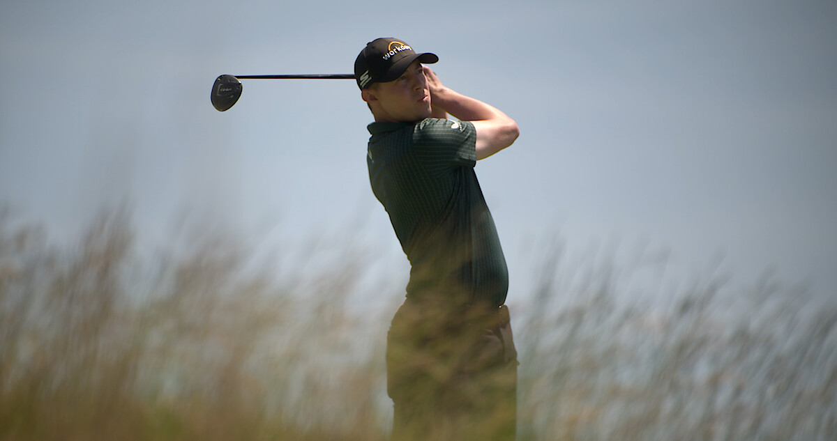 Full Swing Golf Documentary Release Date, Cast, Trailer - Netflix Tudum22 setembro 2024
 -
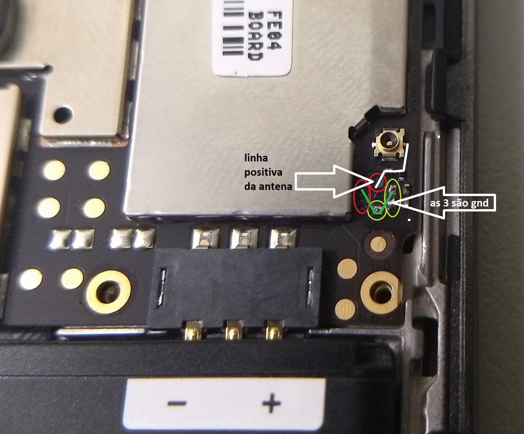 Jumper na antena Moto G4 Play (XT1603) - REPAROS NO HARDWARE - Clan GSM22 setembro 2024
 -
 Anime Expo 2017: Food Wars English Dub Cast Announced - IGN22 setembro 2024
 -
 Oque eu faço, sou mobile e fui banido por nada (Game: Arsenal) roblox* O cara voltando dps de 2099999815 dias - iFunny Brazil22 setembro 2024
 -
 Samsung 43” Class CU7000 Crystal UHD 4K Smart Tizen TV UN43CU7000FXZA - Best Buy22 setembro 2024
 -
![Oficial] Sony revela PS Plus de fevereiro de 2020](https://meups.com.br/wp-content/uploads/2020/01/PlayStation-Plus-Fevereiro-2020.jpg) Oficial] Sony revela PS Plus de fevereiro de 202022 setembro 2024
Oficial] Sony revela PS Plus de fevereiro de 202022 setembro 2024 -
 Instalando Dublagem FIFA 18 - Narração PT-BR (STEAMPUNKS22 setembro 2024
 -
 Wallpaper Lamborghini, Centennial, Lamborghini Centenary LP 770-4, Forza Horizon 3 for mobile and desktop, section игры, resolution 1920x1080 - download22 setembro 2024