Morphology of Leydig cells in the testes after in vivo MCP-1 treatment.
Por um escritor misterioso
Last updated 21 setembro 2024

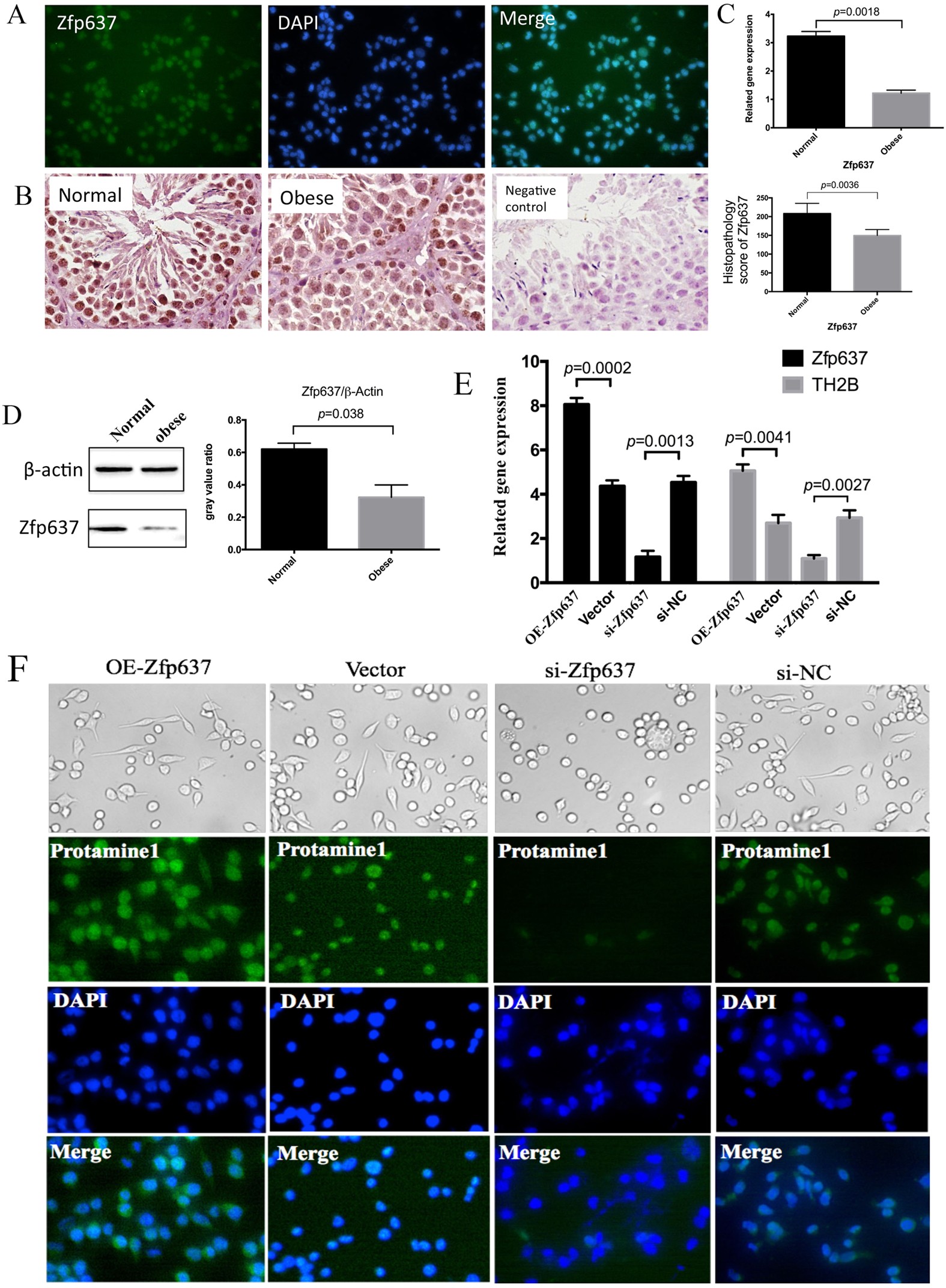
IL-6 mediates differentiation disorder during spermatogenesis in obesity-associated inflammation by affecting the expression of Zfp637 through the SOCS3/STAT3 pathway

Testicular macrophages are recruited during a narrow time window by fetal Sertoli cells to promote organ-specific developmental functions

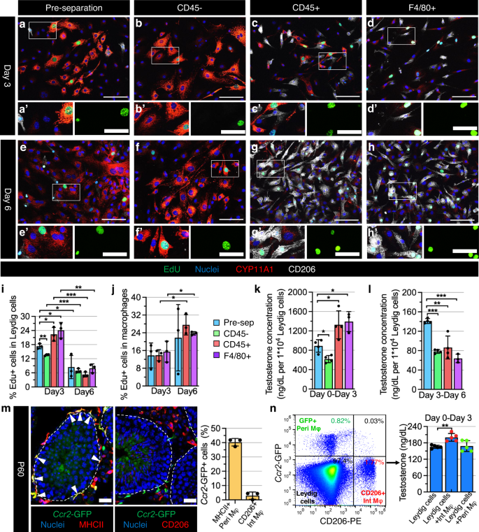
PDF) Monocyte Chemoattractant Protein-1 stimulates the differentiation of rat stem and progenitor Leydig cells during regeneration

A brief exposure to cadmium impairs Leydig cell regeneration in the adult rat testis

Morphology of Leydig cells in the testes after in vivo MCP-1 treatment.

Morphology of Leydig cells in the testes after in vivo MCP-1 treatment.
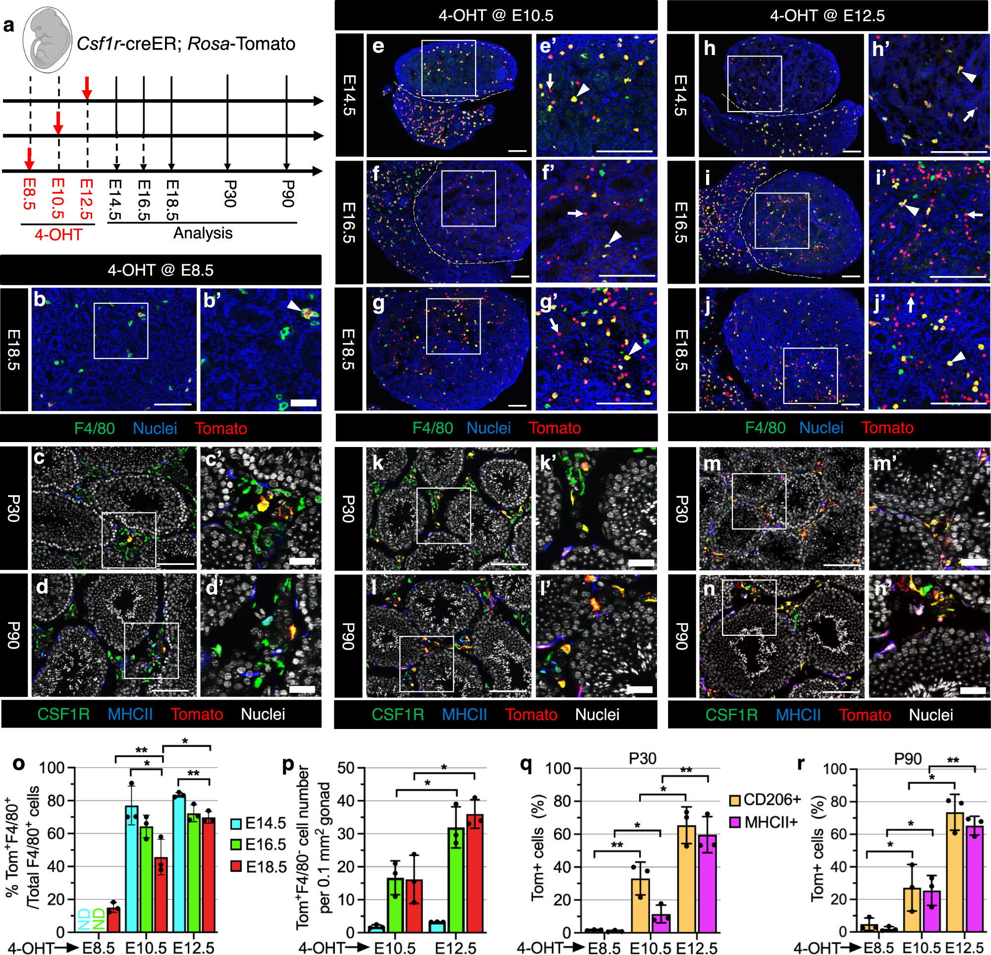
Testicular macrophages are recruited during a narrow fetal time window and promote organ-specific developmental functions
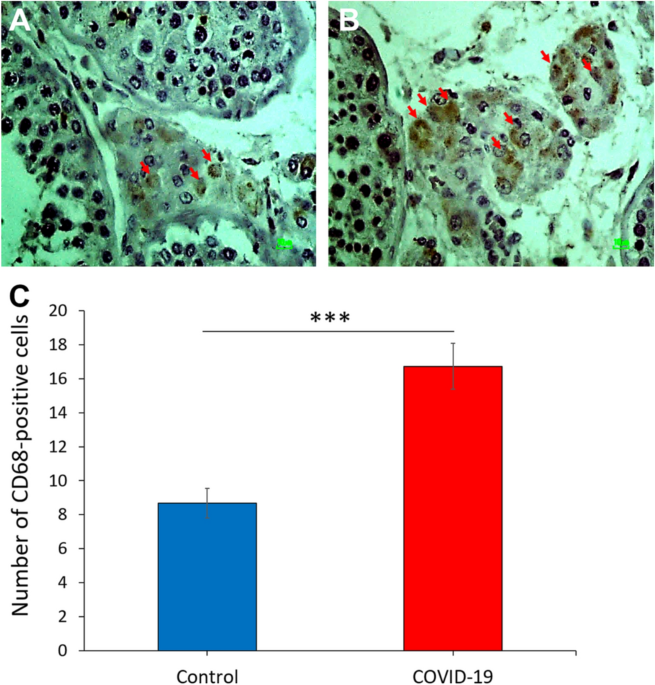
COVID-19 disrupts the blood–testis barrier through the induction of inflammatory cytokines and disruption of junctional proteins
Testicular macrophages are recruited during a narrow fetal time window and promote organ-specific developmental functions

Monocyte Chemoattractant Protein-1 stimulates the differentiation of rat stem and progenitor Leydig cells during regeneration, BMC Developmental Biology

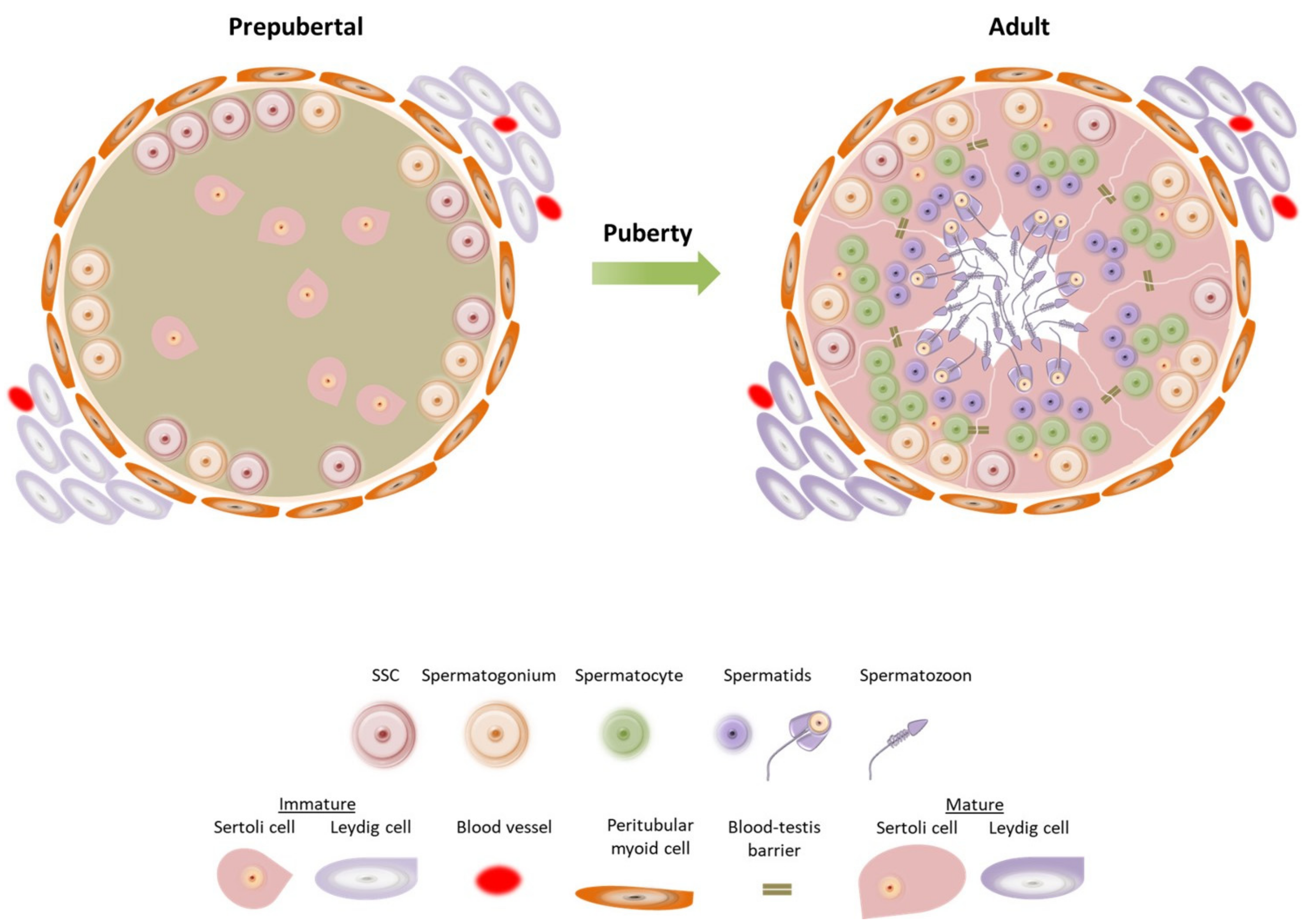
Quantitative Proteomics Reveals the Essential Roles of Stromal Interaction Molecule 1 (STIM1) in the Testicular Cord Formation in Mouse Testis* - Molecular & Cellular Proteomics
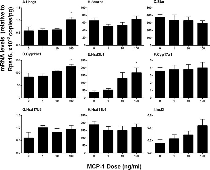
Monocyte Chemoattractant Protein-1 stimulates the differentiation of rat stem and progenitor Leydig cells during regeneration, BMC Developmental Biology
Frontiers Mumps Orchitis: Clinical Aspects and Mechanisms
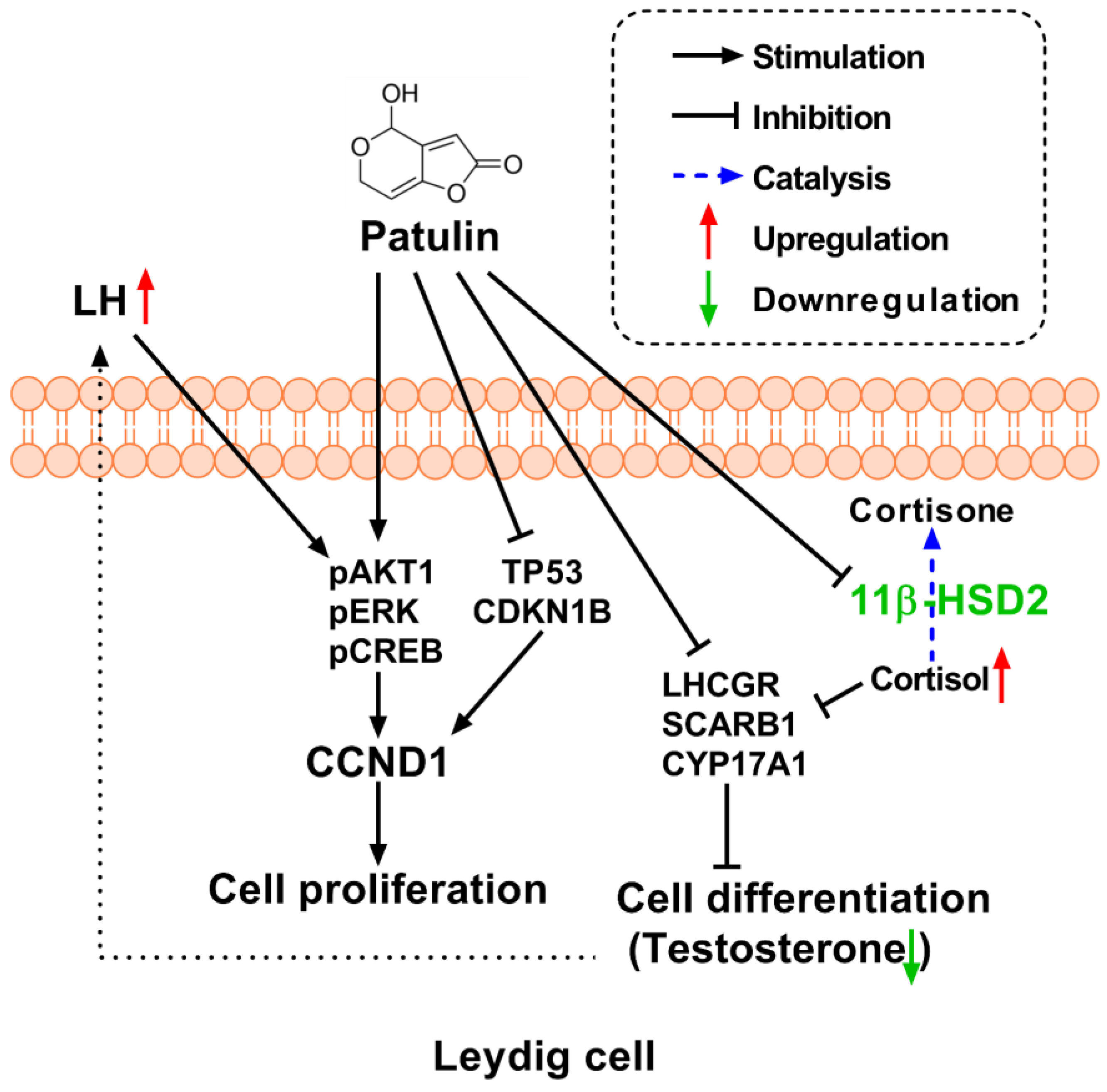
Toxins, Free Full-Text
IJMS, Free Full-Text
Recomendado para você
-
 Teste de Velocidade Vivo, Teste Vivo, Power, Internet21 setembro 2024
 -
 Resultado Teste Vivo Fibra Tv - Vale a Pena?21 setembro 2024
 -
 Domary Avançado RCD testador de soquete elétrico neutro automático circuito de teste de fio terra ao vivo detector de polaridade parede interruptor de tomada ue localizador teste de vazamento elétrico com display21 setembro 2024
 -
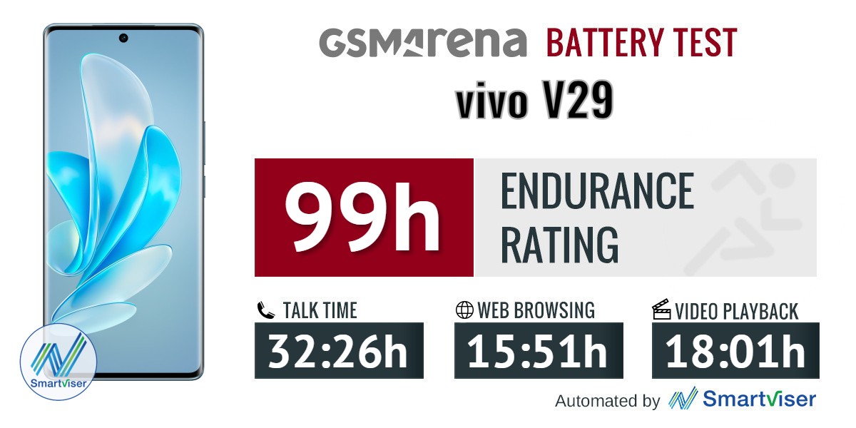 vivo V29 review: Lab tests - display, battery life, charging speed, speaker21 setembro 2024
 -
 Botão webinar ao vivo. ícone de cor azul para curso online, educação a distância, vídeo-aula, conferência em grupo na internet, teste de treinamento. webinar ao vivo com microfone, ícones de transmissão21 setembro 2024
 -
 ROWCES Caneta multímetro digital NCV AC/DC Voltímetro Resistência do ohmímetro Capacitância Frequanecy Teste de linha ao vivo Testador de luz de fundo de LCD portátil de 4000 contagens com retenção21 setembro 2024
 -
![Vivo Fibra 100MB - Teste de velocidade Vivo de 100MB [2018]](https://i.ytimg.com/vi/L-YmHTsWhFc/hqdefault.jpg) Vivo Fibra 100MB - Teste de velocidade Vivo de 100MB [2018]21 setembro 2024
Vivo Fibra 100MB - Teste de velocidade Vivo de 100MB [2018]21 setembro 2024 -
 TESTE GRÁTIS 24 HORAS internet ilimitada da VIVO21 setembro 2024
 -
 Vivo X90 Pro+ Review: Vivo sets the bar very high with its flagship smartphone - Reviews21 setembro 2024
 -
Vivo X60 Pro 5G passa por teste de câmeras e bate Galaxy S21 Plus no ranking do DxOMark21 setembro 2024
você pode gostar
-
 Doodle Dump 8 by ToaPhantom : r/Warframe21 setembro 2024
 -
Deportivo Laferrere - LAFE SE RETIRO DEL MERCADO DE PASES, Y SOLO21 setembro 2024
-
 20 ideias de Tipo Mandrake roupas, moda masculina, moda21 setembro 2024
 -
 Assiste aos primeiro episódios de Pokémon Generations21 setembro 2024
 -
 Dream`s Motel21 setembro 2024
 -
 Casaco Moletom Orlando Magic Canguru Logo - NBA - FIRST DOWN - Produtos Futebol Americano NFL21 setembro 2024
 -
hello kitty pants roblox|TikTok Search21 setembro 2024
-
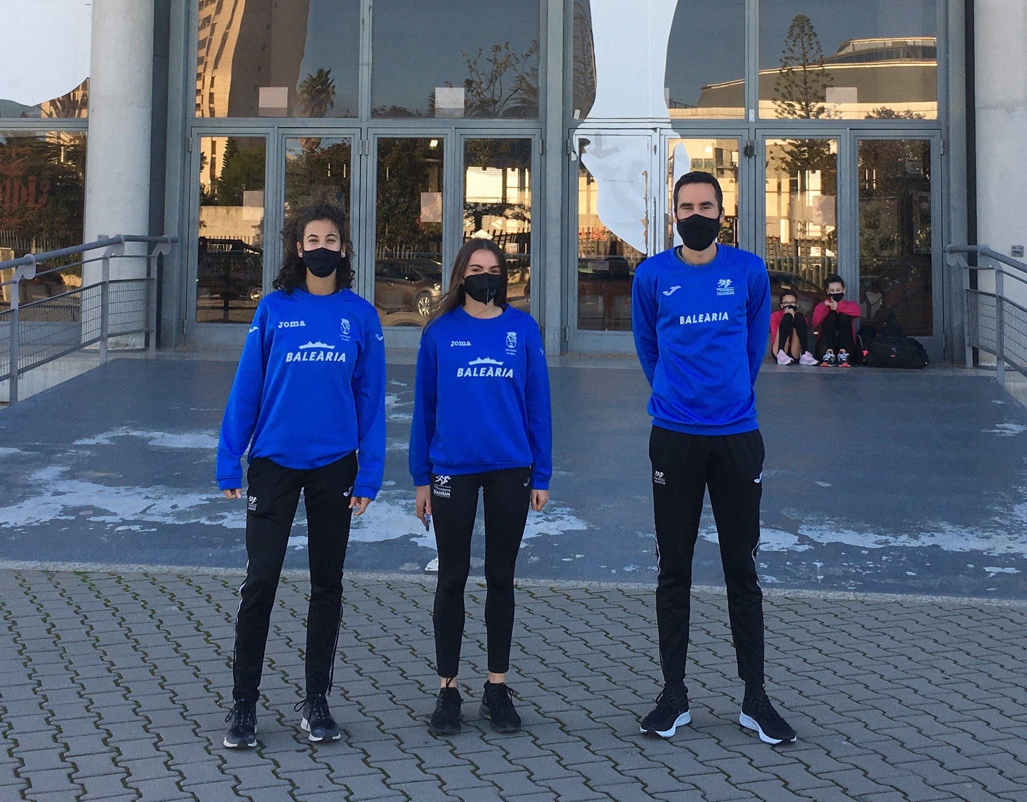 Atletas del CA Baleària Dianium seleccionadas para las concentraciones de la FACV - Dénia.com21 setembro 2024
 -
 Tarot Cigano – Como jogar e significado das cartas21 setembro 2024
 -
 Macaco de pelúcia pet: muita diversão para os cães21 setembro 2024

